Diagnostic lines of defense in cardiovascular health have evolved significantly, allowing physicians to move beyond visualizing the surface of the heart with the stethoscope to actually studying the function and pathology of the cardiovascular system.
The Echocardiogram (ECG) or Color Doppler Imaging is an indispensable tool for a quick assessment of serious cardiac diseases. Working as a unit, they are responsible for tri-dimensional diagnostics and give doctors valuable and exact details about the heart morphology, physiology, and distributive functions.
Now, let’s join in a discussion of what their primary function is and how they help doctors treat cardiac issues.
An Overview – Electrocardiogram (ECG):
An electrocardiogram (ECG) stands as one of the oldest and most fundamental tools in cardiovascular diagnostics. It is an image that captures the heart’s electrical activity which is the process of depolarization and repolarization of cardiac muscles.
In clinical practice, the ECG serves multifaceted purposes:
- Detecting Arrhythmias: Heart function irregularities like atrial fibrillation, ventricular tachycardia, or heart blocks may be promptly established with the usage of an ECG pattern.
- Assessing Conduction Abnormalities: Based on measuring the interrelation between specific ECG signals, which indicate conduction delays like bundle branch block or prolonged QT interval, the diagnosis of the patient can be made.
- Identifying Ischemic Changes: ECG variations, manifested as ST-segment elevation or depression, and T-wave inversions are most commonly associated with myocardial ischemia or infarction, and this information contributes significantly to the early diagnosis of acute coronary syndromes.
As the ECG is straightforward to operate, it remains an invaluable tool due to its rapid result delivery, affordability, and lack of invasiveness.
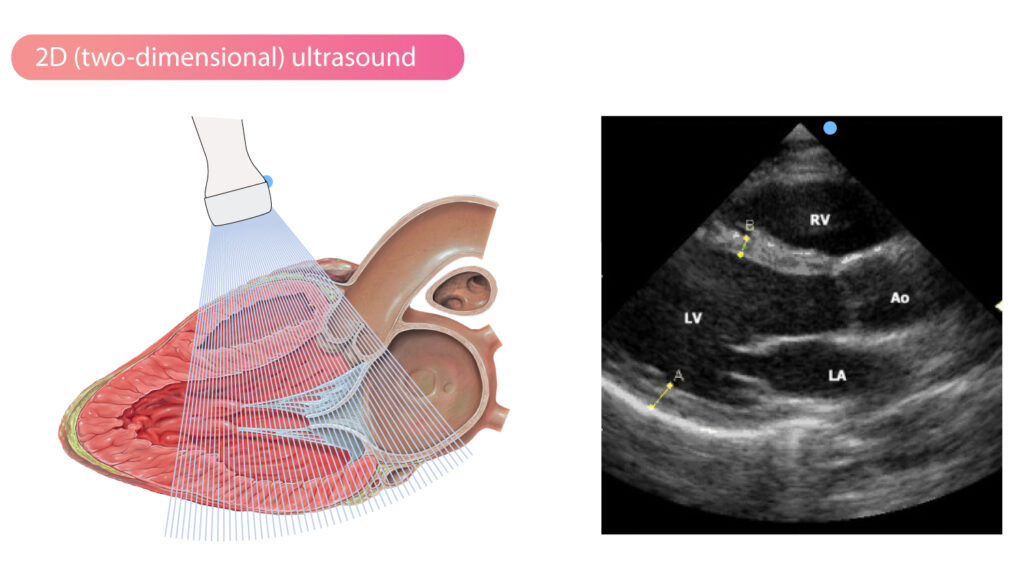
Two-Dimensional Echocardiography (2D Echo):
Two-dimensional echocardiography, or 2D Echo, revolutionized cardiovascular imaging by offering real-time visualization of cardiac structures and dynamics. Utilizing ultrasound waves provides detailed anatomical information, allowing clinicians to assess chamber dimensions, wall motion, and valvular morphology.
The versatility of 2D Echo manifests in various diagnostic realms:
- Structural Assessment: From the assessment of ventricular hypertrophy and dilatation to the detection of congenital heart defects, 2D Echo unveils structural abnormalities with unparalleled clarity.
- Functional Evaluation: By evaluating ejection fraction, wall motion abnormalities, and chamber volumes, 2D Echo aids in gauging cardiac function and diagnosing conditions like heart failure.
- Valvular Assessment: Doppler echocardiography, a subset of 2D Echo, enables precise evaluation of valvular stenosis or regurgitation, guiding therapeutic interventions and surgical planning.
Moreover, advancements like speckle tracking and strain imaging enhance 2D Echo’s sensitivity in detecting subtle myocardial dysfunction, further augmenting its diagnostic utility.
Color Doppler Imaging:
Color Doppler imaging, an adjunct to 2D Echo, elucidates the hemodynamic intricacies of cardiovascular physiology. By superimposing color-coded flow velocities onto anatomical images, it visualizes blood flow patterns, identifying turbulence, shunts, or regurgitant jets.
Key applications of color Doppler imaging include:
- Assessing Valve Function: It facilitates the quantification of valvular regurgitation or stenosis, aiding in severity grading and therapeutic decision-making.
- Detecting Shunts: In congenital heart disease or intracardiac communications, color Doppler imaging delineates abnormal flow patterns, pinpointing the presence and magnitude of shunts.
- Evaluating Pericardial Effusions: By detecting diastolic collapse of cardiac chambers and assessing fluid dynamics within the pericardial space, color Doppler aids in diagnosing pericardial effusions.
Furthermore, spectral Doppler analysis complements color Doppler imaging by providing quantitative measurements of blood flow velocities, enabling the assessment of conditions like diastolic dysfunction or valvular gradients.
All Summed Up!
The immeasurable importance continues to be emphasized as 2D Echo, ECG, and color Doppler imaging techniques become vital in every field of cardiology. Every modality provides a distinct aspect of why one represents electrical activity, other structural characteristics, and cardiac function hemodynamics. Integrating their disciplines, clinicians gain a detailed knowledge of cardiovascular pathology, thus allowing them to choose the right and targeted way of treatment for every patient.
Along with the continued technological advancement and improved diagnostic algorithms, these cardiovascular cornerstone modalities have remained at the center of the medical field’s lineup, driving precision medicine and further making sure improved patient outcomes are achieved. Cardiologists at German Heart Centre tend to gather techniques in the mystery-solving process of the heart, while only care in heart and well-being patients which pay them overall.